Image diagnoses for "skin-colored"
267 results with 583 images
Results forskin-colored


Fox-fordyce's disease L75.2
Fox-Fordyce's disease: 18 years, female: for about 1.5 years axillae bds non-inflammatory papules. relatively symptom-free. anamnesis under steroid cream times been better, after discontinuation recurrence.

Nevus sebaceus Q82.5
Sebaceous nevus: clinical aspect of a sebaceous nevus in a few-month-old infant; only the slight plaque-like elevation of the hairless area indicates the actual diagnosis.


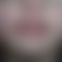
Cheilitis granulomatosa G51.2
Cheilitis granulomtosa: Monosymptomatic orofacial granulomatosis. solitary, chronic, recurrent for months, clearly increased consistency, smooth swelling of the upper lip accompanied by a feeling of tension. no lingua plicata. no facial paresis.

Vascular malformations Q28.88
Malformations vascular. Venousmalformation. Suction. "Angiokeratoma scroti et vulvae".

Birt-hogg-dubé syndrome D23.-
Birt-Hogg-Dubé syndrome: Multiple, skin-coloured, flesh-coloured and whitish, partly waxy, relatively coarse, 2?5 mm large, hemispherical asymptomatic papules retroauricular in a 47-year-old female patient.


Node
Nodules: Keloids : Chronically dynamic, continuously growing for 2 years, hard, red, polypose nodes (keloids in known acne vulgaris).

Aplasia cutis congenita (overview) Q84.81

Graft-versus-host disease chronic L99.2-
Graft-versus-Host Disease, chronic: 1.5 years after stem cell transplantation, large-area scleroderma with significant movement restriction, significant reduction of the AZ

Neurofibromatosis, segmental Q85.0
Neurofibromatosis segmentale: circumscribed soft papules and nodes.

Prurigo simplex acuta L28.22
Prurigo simplex acuta infantum, disseminated, torturously itchy, sometimes excoriated papules and blisters on the right leg of a 10-year-old boy.

Syringome disseminated D23.L
Syringome disseminated:skin-coloured to slightly brownish, completely asymptomatic, surface-smooth, roundish or elongated, broad-based nodules locatedon thetrunk and in the facial region.

Lipoma (overview) D17.0
Lipoma: A subcutaneous lump on the upper arm which has existed for years, is completely unattractive and asymptomatic, can be easily delimited and slides over the underlying tissue.

Onychodystrophia psoriatica

Follicular mucinosis L98.5
Mucinosis follicularis: acute clinical picture developed after heavy sweating; multiple, generalised, 0.1 cm large, itchy, skin-coloured, pointed conical, rough papules bound to follicles.

Asteatotic dermatitis L30.8
Desiccation dermatitis: predominantly coarse lamellar desquamation of the altogether dry skin in the area of the abdomen, caused by treatment with isotretinoin.


Cutis rhomboidalis nuchae L57.2
Cutis rhomboidalis nuchae. coarse-meshed, bulging wrinkles in the neck with massive actinic elastosis. conspicuous follicular prominence with increased retention of the follicular keratin (preliminary stage to the finding in Favre-Racouchaud's disease).