Melanoma amelanotic Images
Go to article Melanoma amelanotic
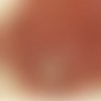
Melanoma malignes, amelanotic: reddish lump that has existed for years and has bled for a few weeks after shaving, otherwise no symptoms.


Melanoma malignes, amelanotic: a reddish lump that has existed for years and bled for the first time a few weeks ago, otherwise no other symptoms.

Melanoma malignes, amelanotic: Overview. reddish lump which has existed for years and bled for the first time a few weeks ago. Otherwise no other symptoms. At the top right side an ulcerated red lump as indication of a metastasis.

Melanoma malignes, amelanotic: Overview. reddish lump which has existed for years and bled for the first time a few weeks ago (encircled). Otherwise no other symptoms. In the upper right corner an ulcerated red lump (square) as indication of a metastasis.

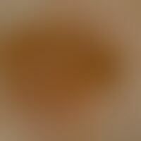
Melanoma malignes, amelanotic: a reddish lump that has developed from a red spot and bled for the first time a few weeks ago. Otherwise no other symptoms.

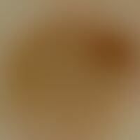
Melanoma malignes, amelanotic: a reddish lump that has existed for years, which has been constantly weeping and bleeding for several months.

Melanoma, malignant, amelanotic. sharply defined, largely amelanotic flat knot. on the right side still distinct melanotic pigmentation. also at the upper rim delicate pigmentation.

Melanoma, malignant, amelanotic. Incident light microscopy. Largely melanin-free parenchyma. Marginal delicate pigmentation, dense in the middle.

Amelanotic lentigo maligna melanoma: regressive originally coherent brown plaque, dissected by degradation processes. For months continuous "decoloration" of the plaques (Inlet: spot-like residual pigmentation marked by arrows). The changes were misinterpreted as "benign development".

melanoma, malignant, amelanotic. for years in the region of the right dorsal lower leg localized (61-year-old man), slowly progressing in size, symptomless plaque measuring 1.5 x 2 cm, with coarsely lamellated scaling. the coloration is mainly red, only focally dark brown. the lower part of the tumor is flat-nodularly raised.


Melanoma, malignant, amelanotic. reflected light microscopy: edge section with discrete pigment strip and adjacent amelanotic tumour.

melanoma malignes amelanotic: since earliest childhood a pigment mark is known at this site. continuous growth for several years. ulceration of the node for half a year. no significant symptoms. the diagnosis cannot be made on the basis of the clinical picture.

Melanoma, malignant, amelanotic. detail enlargement: Cherry-sized tumor, completely eroded on the surface, with yellowish crusts on the edges, sharply defined.

Nodular malignant melanoma (amelanotic). 5 months ago first noticed, cherry-sized, slowly size-progressive, superficially completely eroded, moderately shifting, sharply defined nodule with a reddish to skin-colored coloration (right outer ankle of the foot) in an 83-year-old female patient (TD: 5 mm; Clark level IV, pT4b N0 M0; AJCC IIC).

melanoma malignes amelanotic: since early childhood a pigment mark is known at this site. continuous growth for several years. for half a year extensive ulceration of the node. no significant symptoms.

melanoma malignes amelanotic: since early childhood a pigment mark is known at this site. continuous growth for several years. flat ulceration of the node for half a year. no significant symptoms. arrows mark the non-node parts of the "acrolentiginous melanoma".

Melanoma malignes amelanotic: since early childhood a pigment mark is known at this site. Only in the last months this site has bled with slight traumas. There are no other complaints.

Melanoma, malignant, acrolentiginous. solitary, chronically stationary, slowly increasing, localized at the right big toe, measuring about 0.5 cm, touch-sensitive, red node ulcerated with a dark pigmented part (see circle and arrow marking) Histology: tumor thickness 2.7 mm, Clark level IV, pT3b N0 M0, stage IIB.

Melanoma, malignant, acrolentiginous. solitary, chronically stationary, slowly increasing, localized at the right big toe, measuring approx. 0.5 cm, touch-sensitive, red node ulcerated with a dark pigmented part (see circle and arrow marking) Histology: tumor thickness 2.7 mm, Clark level IV, pT3b N0 M0, stage IIB.