Basal cell carcinoma nodular Images
Go to article Basal cell carcinoma nodular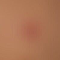


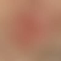
Basal cell carcinoma, solid. chronic, reddish lump with a shiny, smooth surface. clinical and incident light microscopic detection of tumor-specific, bizarrely configured, carmine red vessels extending over the rim wall.

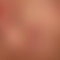
Basal cell carcinoma, solid. solitary, chronically dynamic, 1.2 x 0.8 cm in size, well defined, firm, red ulcer with prominent, shiny marginal wall.

Basal cell carcinoma, solid, sharply defined, slow-growing, approx. 5 mm diameter, smooth, shiny, rough papules.

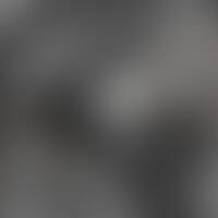
Basal cell carcinoma, solid. reflected light microscopy: In the centre of the figure smaller keratotic plaques surrounded by bizarre, arcade-shaped, irregularly calibrated ?tumour vessels? Histology: solid, undifferentiated basal cell carcinoma.

Basal cell carcinoma, solid. nodular, basaloid tumor formations which penetrate almost the entire dermis. at the base of the tumor dilated eccrine sweat glands. in the picture on the right a cystic dilated hair follicle.

Basal cell carcinoma, solid. intensely basophilic, solid epithelial strands consisting of largely uniform, cytoplasmic-poor, basaloid cells with roundish to oval, partly also spindle, basophilic nuclei. peripheral palisade-like formations. the internal tumor parts are largely unstructured. differentially diagnostically helpful are the cystic-vacuolated cleavages between tumor parenchyma and tumor stroma which always occur in this BCC type.