Image diagnoses for "Nodules (<1cm)", "Face", "skin-colored"
15 results with 25 images
Results forNodules (<1cm)Faceskin-colored
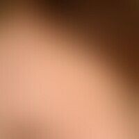
Verrucae planae juveniles B07
Verrucae planae juveniles: Single polygonal, yellowish papules the size of a pinhead on the left forehead of a 10-year-old girl which have been added (inoculation) for weeks.

Nevus melanocytic dermal type D22.L
Dermal melanocytic nevus: for 12 years persistent, 0.9 x 0.9 cm in diameter, soft, sharply defined, calotte-shaped skin-coloured lump on the forehead. 76 year old female patient: "In former times a brownish birthmark had been located at this site".
Multiple Trichoepithelioma D23.-
Trichoepitheliomas: diffusely distributed, small, skin-coloured papules in the forehead area.

Sebaceous gland carcinoma C44.L4
Carcinoma of the sebaceous glands: unspectacular, not spectacular, firm, broadly seated nodule.

Giant cell arteritis M31.6
Arteriitis temporalis. string-like thickened, focal indurated and painful arteria temporalis. at the same time strong, right-sided, temporal headache. no visual disturbances.
Brooke-spiegler syndrome Q85.8
Brooke-Spiegler syndrome: multiple trichoepitheliomas in Brooke-Spiegler syndrome
Contagious mollusc B08.1
Small papular type of Mullusca contagiosa: focal sowing of small papular skin-coloured, smooth efflorescences reminiscent of verrucae planae juveniles; isomorphic irritant effect detectable.
Lymphoepithelioma-like carcinoma C44.4
Lymphoepithelioma-like carcinoma: unspectacular clinical picture with glassy appearing solid nodules. Fig. taken from Oliveira CC et al. (2018) Lymphoepithelioma-like carcinoma of the skin. An Bras Dermatol 93:256-258.
Verrucae planae juveniles B07
Multiple Trichoepithelioma D23.-
Trichoepitheliomas: bulging elastic skin-coloured nodules and nodules in the nasolabial fold; multiple, rough, hemispherical, 0.2-0.5 cm large, partly glassy, skin-coloured or red, symmetrically arranged nodules
Basal cell carcinoma sclerodermiformes C44.L
Epidermal cyst L72.0
Epidermal cysts. 48-year-old female patient, skin change since 1 year, progressive. findings: Multiple, disseminated, localized on forehead and cheeks, skin-colored, rough, noncolored papules with smooth surface, about 0.3 x 0.8 cm in size.
Multiple Trichoepithelioma D23.-
Trichepitheliomas: disseminated, small, firm, symptomless skin-coloured papules in the forehead area.
Nevus melanocytic dermal type D22.L
Dermal melanocytic nevus: known since earliest childhood. Only in recent years clear exophytic growth. The birthmark has become increasingly discoloured and the growing bristle hairs are depilated regularly.
Verrucae planae juveniles B07
Verrucae planae juveniles: Polygonal, yellowish papules the size of a pinhead, partly with discrete scaling, first appeared on the chin of a 10-year-old boy half a year ago.
