Synonym(s)
HistoryThis section has been translated automatically.
Albers 1816
DefinitionThis section has been translated automatically.
Argyrosis (argyria) is a deposition disease caused by chronic exposure or ingestion of silver. A distinction is made between:
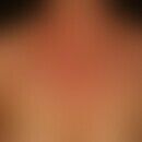
- Generalized argyrosis: Slate-grey, diffuse pigmentation of skin and mucous membranes with accentuation of light-exposed areas due to deposition of silver complexes (oxidation products of the primarily colourless silver complexes: photo effects as in photography) after ingestion of silver-containing drugs.
- Localized argyrosis: circumscribed slate-grey discoloration, especially in the mucous membrane area, after application of preparations containing silver salts. Also after piercing!
You might also be interested in
EtiopathogenesisThis section has been translated automatically.
Ingestion of silver-containing medicines, e.g. silver protein acetyltannate in Targesin roll-on cures, silver compounds in throat disinfectants.
Chronic exposure to silver dust among employees in silver processing plants.
Lozenges for smoking cessation can also contain silver salts.
Silver bound to cysteine enters the skin via the bloodstream. When exposed to light, a photochemical reaction takes place, producing poorly soluble Ag2S(silver sulphide), which accumulates in the skin. (Principle of the exposed photo plate).
Silver also stimulates melanin production, which further intensifies sunlight-triggered hyperpigmentation (Cavdar M et al. 2025)
LocalizationThis section has been translated automatically.
ClinicThis section has been translated automatically.
HistologyThis section has been translated automatically.
Numerous black granules visible only at high magnification with affinity to elastic fibers, basement membrane, sweat and sebaceous glands. Illumination of the embedded particles in the dark field.
Electron microscopy: electron dense, approximately 200 nm granules. X-ray microanalysis can be used to detect their high silver content.
Differential diagnosisThis section has been translated automatically.
Physiological (actinic) skin pigmentation: light as in argyrosis; the slate-grey shiny aspect of the skin discoloration is missing.
Cyanosis: blue-red discoloration; depending on the type of cyanosis, lips and mucous membranes are markedly cyanotic, usually a sign of heart failure.
Chrysiasis: brown discolouration of the skin, accentuated by light! Medication history. Clinically not distinguishable without further explanation.
Hydrargyrosis: used to be used as a bleaching agent. Its use is prohibited.
Icterus: elevation of bilirubin; yellowing of the sclerae; liver diseases (e.g. hepatitis, cirrhosis of the liver)!
Xanthoderma: Yellowing of the skin as a side effect of a mepacrin therapy (anti-epileptic drug) or a therapy with Qinacrin or Sorafenib.
Dyschromia caused by drugs such as: minocycline, amiodarone, imipramine, clofazimine, hydroxychloroquine.
Exogenous dyschromia: discoloration of the contact zones.
TherapyThis section has been translated automatically.
Causal therapy not known.
Analysis and avoidance of silver sources, e.g. silver-containing medication!
After avoiding silver intake, the changes do not or only slightly regress.
Chelating agents or hydroquinone proved to be ineffective, see also Chrysiasis.
Successes with localized argyrosis using Nd:YAG laser or alexandrite laser have been reported (Almurayshid A et al. 2020).
LiteratureThis section has been translated automatically.
- Almurayshid A et al. (2020) Effective laser treatment options for argyria: Review of literatures. J Cosmet Dermatol 19:1877-1882.
- Becher E et al. (1994) Generalized argyria due to a preparation with trace elements. Z Hautkr 69: 388-390
- Cavdar M et al. (2025) A case of argyria as consequence of alternative medical treatment. J Dtsch Dermatol Ges 23: 871-873.
- Fisher NM et al. (2003) Scar-localized argyria secondary to silver sulfadiazine cream. J Am Acad Dermatol 49: 730-732
- Fritsch P (1996) Metal dermatoses II. Dermatologist 47: 400-409
- Mittag H et al. (1987) On the question of argyria. Dermatologist 38: 670-677
- Robinson-Bostom L et al (2002) Localized argyria with pseudo-ochronosis. J Am Acad Dermatol 46: 222-227
- White JM et al. (2003) Severe generalized argyria secondary to ingestion of colloidal silver protein. Clin Exp Dermatol 28: 254-256
Recommended articles


. plaque-shaped...")

Incoming links (9)
Addison's disease; Argyrose; Chromonychia; Chrysiasis; Cutis aurantiasis; Dyschromia; Silver; Spot darker; Stain;Outgoing links (10)
Alexandrite laser; Chrysiasis; Cyanosis; Dyschromia; Hydrargyrosis; Hydroquinone; Icterus; Neodymium yag laser; Pigmentation, actinic; Xanthoderma;Disclaimer
Please ask your physician for a reliable diagnosis. This website is only meant as a reference.


: localized...")