Nevus spitz Images
Go to article Nevus spitz
Naevus Spitz: a constantly growing, slightly raised, irregularly bordered, black-brown neoplasm, appearing already in the first months of life, in a now 3-year-old child.
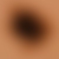
Nevus Spitz: reflected light microscopy of the previously clinically imaged nevus. irregular pigmentation with unstructured, irregularly bordered black center. isolated pseudopodia. marginal reticular pattern.
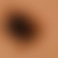
Nevus Spitz: Incident light microscopy of the previously clinically imaged nevus. Irregular pigmentation with unstructured, irregularly bordered black center (arrow) Isolated pseudopodia (rectangles). Random reticular pattern (circles).
Naevus Spitz: slightly raised, irregularly bordered, black-brown neoplasm, existing for several months, in a 4-year-old child.
Naevus Spitz: reflected light microscopic image with a homogeneous, black-brown centre and numerous marginal pseudopodia.
Naevus Spitz: a slightly raised, sharply defined, irregularly pigmented tumour that has existed for several months.

Naevus Spitz: Incident light microscopy of the tumor clinically shown above; irregular pigmentation; black dots.
Naevus Spitz: a brown plaque that has existed for several months, flatly protuberant, sharply defined, irregularly pigmented, completely non-irritant.
Nevus Spitz. reflected light microscopy of the previously clinically imaged nevus. irregular pigmentation; radial-strictive basic architecture, which is particularly visible at the edges, no vascular polymorphies.

Naevus Spitz: rapidly growing, irregularly pigmented tumor on the knee of a 5-year-old girl.

Nevus Spitz: Incident light microscopy (Junction-type epithelial cell nevus): Spot-shaped, centropapillary capillary capillary vessel ectasia distributed over the entire lesion, surrounded by light brown in a quadrant of dark brown melanin pigment.

Nevus Spitz: symmetrical melanocytic compound type nevus, sharply defined towards depth.

Naevus Spitz: Detail enlargement: Hyperplastic epidermis, pronounced ortho-hyperkeratosis. intraepithelial and dermal nests with spindle-shaped melanocytes. characteristic cleavage between epithelium and melanocyte nests. loose round cell infiltrates in the dermis.

Naevus Spitz: Compound type: Epithelial hyperplasia, irregular pigmentation; large nests with irregularly pigmented, predominantly spindle-shaped melanocytes in the epidermis; clumpy melanophagus layer in the dermis

Naevus Spitz: Detail enlargement: Intraepithelial nests of spindly, clumpy pigmented melanocytes. Strong pigmentation of the epithelium. Gap formation between melanocyte nest and epithelium.

Nevus Spitz: dermal melanocytic nevus with bizarrely configured epithelioid melanocytes. distinct dermal fibrosis. no junctional activity.

Naevus Spitz: Detail section: Bizarre configuration of melanocytes, which show partly spindle and partly epithelioid structures, with large, extremely polymorphic nuclei; presentation of the S100 antigen.