Nevus melanocytic (overview) Images
Go to article Nevus melanocytic (overview)
Nevus, melanocytic. Type: Congential melanocytic nevus.

Nevus, melanocytic. Type: Congential melanocytic nevus.

Nevus, melanocytic. type: Congenital melanocytic nevus. solitary, chronically stationary, constant size for 12 months, indolent, sharply defined, axisymmetric, 0.6 cm diameter, dark brown, smooth nodule.
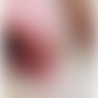

Nevus, melanocytic. Type: Congential melanocytic nevus.

Nevus, melanocytic. Type: Congential melanocytic nevus. Incident light microscopy.

Naevus melanocytic: Large congenital completely unattractive melanocyticnaevus.

Several acquired melanocytic nevi in a 65 years old man. Especially the two conspicuous large melanocytic nevi did not show any changes in the last years. There is no need for surgical removal. Annual clinical controls are necessary.

Nevus, melanocytic, type: Congenital melanocytic nevus.

Nevus, melanocytic. Type: Acral, acquired melanocytic nevus.

Nevus, melanocytic. Type: Congential melanocytic nevus.

Usual melanocytic nevus. Chronic stationary, 0.2-2.0 cm in size, sharply defined, soft, light to dark brown, indolent, smooth papules and plaques in the breast area of a 22-year-old woman. No increase in pigmentation or size is noticeable.

Usual melanocytic nevus. Brownish, roundish, 0.3 cm in diameter, sharply defined, soft, asymptomatic, smooth papule in the region of the right areola of a 26-year-old woman. No size growth in recent years.

Common melanocytic nevus. sharply defined, acquired, homogeneously pigmented melanocytic nevus. surface relief and the hair follicles are preserved

Usual melanocytic nevus. type Lentigo solaris. Chronic stationary, no longer increasing, sharply limited, symptomless, axisymmetric, 1.0 x 0.6 cm large, brown spot on the auricle of a 36-year-old woman.

Nevus, melanocytic. type: Acquired dysplastic melanocytic nevus. solitary, chronically inpatient, approx. 0.7 cm high, light accentuated spot localized at the right temple, smooth, reticularly decomposed with differently graded brown tones, blurredly limited in a 50-year-old female patient.

Nevus, melanocytic. type: Acquired dysplastic melanocytic nevus. Detail: The reticular, dark brown structure of the melanocytic compound nevus is clearly visible. Multiple, oval, partly confluent, round, light brown areas penetrate the nevus. In the center right gray fog zone as a sign of inflammatory infiltration.

Nevus, melanocytic type: Dysplastic melanoytic nevus, growth in the last 6 months, asymmetry of the tumor and the different coloration should in this case lead to an excision and histopathological examination to exclude malignancy (malignant melanoma).


Nevus, melanocytic. Congenital melanocytic nevus of the spilus nevus type

Common melanocytic nevus. dermal type. acquired, known for decades, solitary, chronically inpatient, 0.4 cm tall, sharply defined, soft, symptomless, skin-colored, smooth papules. follicular openings are visible.

Naevus melanocytic (Naevus Spitz.), existing for several months, flatly protuberant, sharply defined, irregularly pigmented, completely non-irritant, brown plaque, 5-year-old boy.

Nevus melanocytic nevus Spitz reflected light microscopy.

Common melanocytic nevus. type: Halo-nevus, almost complete regression of the melanocytic nevi, which are indicated as light brown spots in the middle of the pigment-less areas.

common melanocytic nevus. type: nonfamilial syndrome of (acquired) dysplastic melanocytic nevi. up to 0.5 cm in size, brown, soft papules with smooth surface in disseminated distribution on the entire trunk in a 29-year-old patient. since earliest childhood strong sun exposure during regular bathing holidays at the north sea. the moles "have always been".

Nevus melanocytic. Type: Giant congenital nevus of the swimsuit type.

Congential melanocytic nevus as a cutaneous mosaic.

Melanocytic nevus. Type: Congenital melanocytic nevus. Repigmented scar after partial resection.


Melanocyte nevus, reflected light microscopy

Melanocytes: normal skin; typical physiological position of spindle or star-shaped melanocytes in the basal stratum of the epidermis.

Usual melanocytic nevus. dermal type. nodular new formation with diffuse spreading of melanocytes in the upper and middle dermis. lateral to the nevus intact hair follicles. at the base 2 sebaceous gland complexes.

Common melanocytic nevus, verrucous melanocytic nevus of the dermal type, with a sharply defined tumor parenchaym at the base.


nevus, melanocytic. detail enlargement: melanocytes in the upper dermis in nests, in the middle and deep dermis diffuse and arranged in strands. no melanocyte nests in the epithelium.

Usual melanocytic nevus, junctional type. detail: irregular elongation of the reteleal ridges with diffuse penetration by melanocytes (light elements). in the picture left nest formation of melanocytes at the lower pole of the reteleal ridge. in the dermis a thin lymphocytic infiltrate; isolated melanophages.

nevus, melanocytic. junctional type. elongation of the reteleases. increased basal pimentation. nests of melanocytes at the tips of the reteleases. confluence of the reteleases at several sites (e.g. right image section) of the section.

common melanocytic nevus, junctional type. irregular broadening of the epithelium. orthokeratosis. in the basal epithelial sections and the reteleal ridges there are large nests with melanocytes. in the melanocytes there are medium-sized, roundish, moderately basophilic nuclei. distinctly pronounced cytoplasmic border. partly fine-granular, partly clumpy pigmentation. in the upper dermis numerous melanophages with fine-grained or clumpy pigment.


Melanocyte nevus, laser scanning microscopy, homogeneous junctional nests, overview

Melanocyte nevus, laser scanning microscopy, homogeneous junctional nests, detailed view