HistoryThis section has been translated automatically.
The first description of cockades dates back to 1867 by v. Hebra, who described the cockades of erythema exsudativum multiforme as symmetrically distributed lesions on the back of the hands and feet, forearms and thighs with relative exclusion of the head and trunk. He usually observed the first efflorescences on the back of the hand. The efflorescences were initially lentil- to bean-sized papules that resembled chilblains. In the course of time, the lesions blew off centrally. A second, concentric ring then forms. The skin lesions healed, leaving behind a coarse scaling and slight pigmentation (post-inflammatory pigment incontinence). Von Hebra named the disease "erythema exsudativum multiforme" because, according to his observations, the efflorescences were very variable during development and healing.
DefinitionThis section has been translated automatically.
You might also be interested in

ClassificationThis section has been translated automatically.
For non-autoimmunologic bullous diseases, the type of cockade formation and the extent of the lesional skin changes are now regarded as decisive criteria for their classification. Although the authors describe the extent and localization of mucosal involvement as an important inclusion or exclusion criterion in the SJS/TEN and EEMM disease spectrum, it does not serve to differentiate between the disease entities, as mucosal involvement can be equally severe in the various diseases. Even dermatohistopathological findings do not allow a reliable distinction to be made, as complete epidermal necrosis may be present histologically in all entities. The definition and thus differentiation of the two diseases was therefore based on the criteria
- "Pattern of cocardial lesions"
- and
- "extent of the skin detachment as a percentage of the body surface".
Four different types of cockade lesions were observed and described:
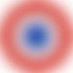
- Typical cockades: Defined as single, regular, round lesions up to three centimeters in diameter. Three zones can be recognized on the typical cockade: a central blister, a raised, edematous central ring and an erythematous outer ring. In the literature, typical cockades are sometimes also referred to as "shooting target lesions" or "iris lesions".
- Raised atypical cockades: Round, palpable, edematous lesions with only two zones and a blurred border.
- Flat atypical cockades: Round lesions with only two zones and a blurred border. With the exception of the central blister, the flat cockades are not raised.
- Figured (targetoid) erythema with or without blistering: red round patches of irregular demarcation, shape and size, often confluent and not palpable (Bastuji-Garin et al. 1993).
LiteratureThis section has been translated automatically.
Bastuji-Garin S et al. (1993) Clinical classification of cases of toxic epidermal necrolysis, Stevens-Johnson syndrome, and erythema multiforme. Arch Dermatol 129: 92-6.
Martin SR (2023) Clinic, epidemiology and etiology of erythema exsudativum multiforme majus Inaugural dissertation for the award of the medical doctorate of the Medical Faculty of the Albert-Ludwigs-University Freiburg im Breisgau
von Hebra F, Kaposi M (1860) Acute exanthema and skin diseases. Textbook of skin diseases. Volume 1, Enke, Erlangen
- von Hebra, FR (1867). Erythema Exsudativum Multiforme (Hebra), in: Atlas der Hautkrankheiten. Published by Ferdinand Enke, pp. 3-4.
Recommended articles


Outgoing links (1)
Erythema multiforme, minus-type;Disclaimer
Please ask your physician for a reliable diagnosis. This website is only meant as a reference.