Cherry angioma Images
Go to article Cherry angioma


Angioma, senile. reflected-light microscopy: sharply defined, partly isolated (marginal area), partly aggregated (in the centre) lobules; unaffected skin with an inconsistent reticular pigmentation pattern and bizarre, completely pigment-less areas (fair-skinned sun-sensitive patient!).

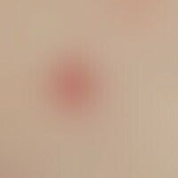
Angioma senile. red brown, very soft papules, almost completely compressible by finger pressure, 0.7 cm in size. therapy not necessary

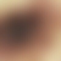
Angioma, senile. 55 years old female patient, in whom this finding has existed for two years. Size progressive, soft, spongy, flat raised, 0.8 x 0.6 cm large lump with a fielded surface.

angioma, senile. 7 mm large lump on the cheek of a 70-year-old patient, existing for years, reddish-brown, very soft, almost completely compressible by finger pressure. skin clearly light-damaged; above left numerous linear telangiectasias. therapy not necessary; if necessary excision without safety distance.

Angioma, senile, multiple, bright red, persistent, hardly increasing in size, disseminated standing papules; the angiomas have been present in the patient for more than 10 years.

Angioma, senile. multipe, chronically stationary,1-4 mm large, sharply defined, initially light red, later dark red to violet, soft, flat papules. patient reported severe seborrhea on the integument.

Angioma, senile. Multipe, chronic stationary, disseminated, erythematous, soft papules

Angioma, senile. Multipe, chronic inpatient, disseminated, erythematous, soft papules in a 70-year-old man.


Angioma, senile. detail enlargement; slight irregular acanthosis of the epithelium; orthokeratosis; convolute thin-walled, ectatic vessels bulging with erythrocytes; the sinus-like cavities in the vascular lumina correspond to shrinkage artifacts.